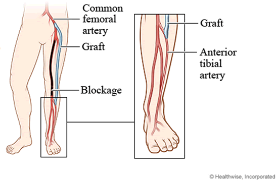
A peripheral vascular bypass, also called a lower extremity bypass, is the surgical rerouting of blood flow around an obstructed artery that supplies blood to the legs and feet.
This surgery is performed when the buildup of fatty deposits (plaque) in an artery has blocked the normal flow of blood that carries oxygen and nutrients to the lower extremities. Bypass surgery reroutes blood from above the obstructed portion of an artery to another vessel below the obstruction. Bypass grafts can be prosthetic material or vein.
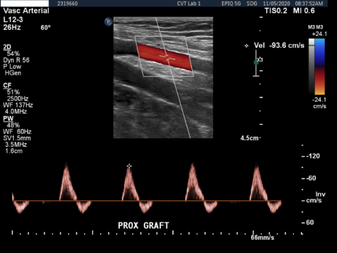
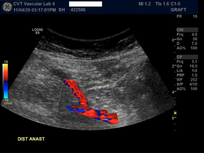
Ultrasound evaluation easily detects a narrowing that can subsequently develop within the graft and allows for repair before the graft is completely blocked.
Bypass grafts are evaluated at three-month intervals for the first year after surgery. Surveillance studies are done every six months in the second year, then annually thereafter. Shorter interval surveillance may be recommended if an irregularity is found.
In addition to a complete ultrasound scan of the graft, ankle, and arm blood pressures will be measured using inflatable cuffs.
Treatment is usually needed if a Deep Vein Thrombosis (DVT) is found. In most cases, this involves treatment with blood thinners.
Length of exam: 45-60 minutes
Preparation: No special preparation is required.
Images of a Bypass Graft Examination